Thermo Fisher Scientific › Electron Microscopy › Electron Microscopes › 3D Visualization, Analysis and EM Software › Use Case Gallery
Mammalian epididymal epithelial cells are crucial for sperm maturation. Historically, vacuole-like ultrastructures in epididymal epithelial cells were observed via transmission electron microscopy but were undefined. Here, we utilize volume electron microscopy (vEM) to generate 3D reconstructions of epididymal epithelial cells and identify these vacuoles as intercellular organelle reservoirs (IORs) in the lateral intercellular space (LIS), which contains protein aggregates, autophagosomes, lysosome-related organelles and mitochondrial residues. Immunolabelling of organelle markers such as P62, LC3, LAMP1 and TOMM20 confirm these findings. The IOR size or number varies across four epididymal regions and decreases with age. Rab27a mutant mice exhibit reduced IORs in the caput epididymis and a subfertility phenotype, suggesting the involvement of Rab27a in the formation of IORs. Furthermore, we observe the presence of IORs between intestinal epithelial cells besides epididymis. Amino acid transporters at IOR edges suggest dynamic protein recycling. Our findings reveal that the IOR is an important structure critical for organelle turnover and recycling outside epithelial cells with limited selfdegradation capabilities.
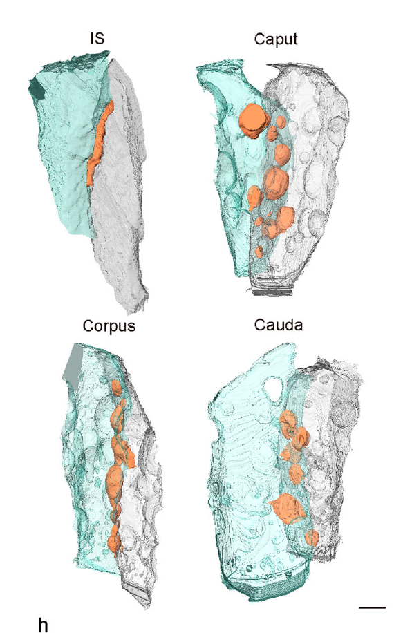
We individually segmented the cell membrane and vacuole membrane via Amira software. […], All 3D segmentations were generated via Amira software (Version 6.5.0, ThermoFisher,USA). RawTiff images (1300 ~ 1500/sample)were imported into Amira for alignment […], Direct 3D reconstruction, volume rendering and visualization of the nucleus, cellmembranes, and IOR were conducted manually upon careful visual inspection with Amira software (Version 6.5.0, Thermo Fisher, USA). […], IORs containing autophagy-like structures were segmented with subvolume extraction created in Amira.
For Research Use Only. Not for use in diagnostic procedures.